Sickle Cell Disease (SCD) and Blood Transfusion: Prevalence of Alloantibodies and Red Cell Antigens among Multiply Transfused Individuals with SCD in Selected Tertiary Health Institutions in Abuja, Nigeria
 , Adeola Olanrewaju Oluboyo PhD1, Celine Ijeoma Okolo BMLS3, Akinloye Bankole Oyetunde MBA, MSc, MPM1, Ifeoma Evelyn Ezeukwu MBBS2

*Corresponding author: Chisara Sylvestina Okolo, Department of Medical Laboratory Sciences, Afe Babalola University, Ado, Ekiti, Nigeria. chisara2001@yahoo.com
-
Received: ,
Accepted: ,
Abstract
Background and Objectives:
Most individuals with Sickle cell disease (SCD) are candidates for frequent blood transfusions, and the chances of alloantibodies production are very high. SCD is associated with severe morbidity, impaired quality of life, and premature mortality. Some SCD individuals are relatively stable and rarely require a blood transfusion; others often require multiple blood transfusions. This study aimed to determine the patterns of alloantibodies and red cell antigen phenotype frequencies among multiple-transfused SCD individuals at tertiary health institutions in Abuja. The study objectives include determining the distribution of minor red cell antigens in sickle cell individuals, relative to control subjects and to determine the presence and type of alloantibodies in sickle cell individuals and control subjects.
Methods:
The study population consisted of 100 SCD individuals who had at least three transfusions and 100 non-SCD individuals who had not been transfused before as controls. An antibody screening panel (3 cells) and an identification panel (10 cells) from the national health service (NHS) (UK) were used to screen and identify alloantibodies through the tube method in low-ionic-strength solution, albumin, and the anti-human globulin (AHG) phase according to the manufacturer’s instructions. Red cell antigen typing was determined using standard tube techniques for ABO, RH, MNSs, Duffy, and Kidd.
Results:
According to the results obtained from the alloantibody assay, out of the 100 SCD individuals, 16% (4) developed anti-D antibodies, 32% (8) developed anti-E, and 24% (6) developed anti-Fya. Only 4% each developed JKb, Lua, M, Lea, and C. Two percent of the individuals developed anti-S antibodies, while all the control samples were negative. The sociodemographic characteristics revealed that the age range of the subjects was between 1 and 46 years, and the majority were aged >1–10 years (38%). The distribution of the ABO blood groups revealed that 55% were in group O, 15% were in group A, and 19% were in group B. 92%, 50%, 41%, 63%, and 87% of the study population were found to be composed of RhD, RhC, RhE, Rhc, and Rhe, respectively. The incidences of M, Leb, K, and K positivity were 58%, 36%, 39% and 41%, respectively.
Conclusion and Implication for Translation:
Identifying alloantibodies in SCD individuals is crucial, as it can have implications for future transfusions and impact the overall management of the disease. Implementing extended RBC phenotyping for all SCD individuals, especially those receiving regular transfusions, could allow for a more individualized and targeted approach to blood transfusion management.
Keywords
Alloantibodies
Alloimmunization
Sickle Cell Disease
Transfusion
Transfusion Safety
INTRODUCTION
Sickle cell disease (SCD) is the most common severe inherited disease in the world and one of the 50 most common causes of non-communicable death globally.[1] SCD is an inherited hemoglobinopathy associated with significant morbidity, reduced quality of life, and premature mortality.[2,3] It affects an estimated 20–25 million individuals globally, with the highest prevalence observed in tropical regions, particularly sub-Saharan Africa, India, and the Middle East.[4] Nigeria plays a significant role in the global burden of SCD, with an estimated 100,000–150,000 newborns affected each year— representing roughly one-third of the global total.[5]
The prevalence of SCD within Nigeria ranges from 2% to 3%.[6] Some SCD individuals are relatively stable and rarely require a blood transfusion;[7] others often require multiple blood transfusions, which may result in red blood cell (RBC) alloimmunization. Alloimmunization in SCD has several adverse effects, including a markedly elevated risk of hemolytic transfusion reactions, which may include hyperhemolytic reactions and iron overload.[8] The occurrence rate of alloimmunization to red cell antigens is a serious complication of transfusion. Alloimmunization is a well-known risk factor associated with repeated transfusions, and the use of extended RBC antigen matching has been well documented to reduce alloimmunization.[9,10] The most serious consequence of alloimmunization is the risk of developing a delayed hemolytic transfusion reaction (DHTR), which may be grievous. In several DHTR individuals, the hemoglobin level falls below the pre-transfusion level, suggesting that hemolysis of the transfused RBCs and the patient’s RBCs occurred, a condition called hyperhemolysis. In cases of DHTRs, additional transfusions may aggravate hemolysis, further worsening anemia and complicating patient management.[11] This underscores Nigeria’s pivotal role in understanding and mitigating the global burden of SCD. In cases of DHTRs, the destruction of the patient’s RBCs is partly attributed to the development of autoantibodies, often precipitated by prior alloimmunization. It is partially explained by the presence of autoantibodies because alloimmunization is thought to trigger antibody production. However, DHTR/ hyperhemolysis cases also occur due to the absence of detectable alloantibodies or autoantibodies.[8] In African populations, especially in Nigeria, the antigen frequency data are sparse, leading to mismatched transfusions and higher rates of alloimmunization. Furthermore, SCD patients are at high risk of developing alloantibodies due to repeated transfusions. These alloantibodies can complicate future transfusions, making it difficult to find compatible blood, and may lead to hemolytic[12] transfusion reactions, both immediate and delayed. It can impact clinical outcomes, increasing morbidity and hospital stays. Identifying the types of alloantibodies prevalent in SCD patients helps blood banks prepare antigen-negative units and adopt preventive strategies like extended phenotype matching.
The study objectives include determining the distribution of minor red cell antigens in sickle cell individuals and control subjects, and determining the presence and type of alloantibodies in sickle cell individuals and control subjects.
METHODS
This was a cross-sectional study using a purposive sampling method. The study was conducted among individuals with SCD attending the National Hospital Central Area, Abuja, Federal Medical Center Jabi, Abuja, and University of Abuja Teaching Hospital, Gwagwalada, Abuja. The study population included 100 SCD individuals of all ages who had undergone at least three transfusions and 100 healthy non-SCD individuals who had not been transfused before as controls. Quantitative variables were grouped into categories to facilitate comparison between subgroups.
Both qualitative and quantitative methods were used for the data collection and laboratory analysis of the samples. The data were collected over a period of 9 months from October 2022 to July 2023.
This study adhered to the Standards for Reporting Qualitative Research to ensure transparency and completeness in presenting qualitative aspects such as problem formulation, research questions, data collection, and analysis. For components involving numerical data, the Strengthening the Reporting of Observational Studies in Epidemiology guidelines were followed to maintain rigor in methodology, statistical analysis, and presentation of results.[12]
Data Collection
Structured informed consent and assent forms for the minors were provided to the individuals before sample collection was carried out. The individuals were not coerced into the study; they were assured of the confidentiality of their results and that it was a voluntary, non-remuneration exercise. A structured questionnaire was administered to the individuals before sample collection to ensure qualitative and quantitative data collection, and sociodemographic characteristics such as age, sex, and number of siblings.
The procedures in this study followed the principles of Helsinki Declarations and Ethical approval were obtained from the Health Research Ethics Committee of the College of Medicine and Health Sciences, Afe Babalola University Ado Ekiti, Ekiti State, Nigeria (ABUADHREC/12/06/2021/056); National Hospital, Abuja, Nigeria (NHA/EC/083/2022); Federal Medical Center Jabi, Abuja, Nigeria (FMCABJREC/ HREC/2022/076); and University of Abuja Teaching Hospital Gwagwalada, Abuja, Nigeria (FCT/UATH/HREC/1085).
Sample Size and Sampling Technique
The sample size was determined using the sample size calculation, N = (z2 p [1−p])/e2 and based on the prevalence of SCD, which is 3%.
Specifically, N was the minimum sample size.
Where:
Z = confidence level 95% = 1.96 n = Sample Size P = Prevalence rate of SCD 3% = 0.03 e = Error margin accepted = 5% = 0.05.
A total of 45 samples were calculated, but 100 samples were collected from SCD individuals from the facilities mentioned. Another age-matched 100 samples from HB AS and HB AA individuals who have not been transfused before serve as controls.
Inclusion Criteria
Male and female SCD individuals of all ages who had been transfused at least three times were recruited for this study as subjects. Apparently healthy individuals with hemoglobin genotype AA and AS who gave their consent were recruited as control subjects.
Exclusion Criteria
SCD individuals who refused to give written consent/assent, those who have not been transfused before, those transfused in the past 3 weeks, and pregnant women were excluded.
Study Population
A total of 200 blood samples were collected from different sites. One hundred SCD individuals (HB SS, SC, and CC) who had received three or more units of blood and 100 controls (HB AA and HB AS) who had never been transfused were included. Five milliliters of intravenous blood was collected from each volunteer into Becton Dickinson Ethylenediaminetetraacetic acid (BD EDTA) vacutainer tubes.
Variables
In this study, the independent variable was the transfusion status of participants (multiply transfused SCD patients against the non-transfused controls). The dependent variables were the presence and specificity of red cell alloantibodies and the distribution of selected minor red cell antigens. Age, sex, study site, and laboratory techniques were treated as control variables to minimize confounders. Descriptive statistics were employed to summarize the baseline characteristics of the study population. Categorical variables such as sex, presence or absence of alloantibodies, and red cell antigen phenotypes were presented as frequencies and percentages. Continuous variables such as age and number of transfusions were reported as means ± standard deviations (SD).
Antibody Screening
An antibody screening panel (3 cells) and identification panel (10 cells) from NHS (UK) were used to screen and identify alloantibodies through the tube method in low-ionic-strength solution, albumin, and AHG according to the manufacturer’s instructions (3-cell antibody screen in Alsevers-Lot No: R1213671 and 10-cell identification Panel 1 in Alsevers-Lot No: R1443538).
This was aimed at identifying the presence of antibodies in a patient’s blood serum. Indirect antiglobulin tests were performed to detect the presence of unexpected antibodies in the patient’s plasma. Plasma samples were incubated with reagent red cells at room temperature and 37°C to determine the presence of agglutinins. Agglutination confirms the presence of an alloantibody in the plasma.
Antibody Identiflcation
When antibodies were detected, further testing is necessary to identify the specific antibodies present through panel testing: A panel of known RBC antigens and patient plasma were incubated at room temperature, albumin, low ionic saline, and antihuman globulin to determine the specificity of the detected antibodies.
Procedure
Two volumes of test plasma were dispensed into a labeled test tube. One volume of the supplied red cell was dispensed into the test tube and mixed thoroughly. It was then incubated in a water bath at 37°C for 45 min. After incubation, the cells were washed 3 times using normal saline. Two volumes of anti-human globulin reagent were added to the cells. The cells were spun for 3 min, and agglutination was observed. Agglutinated samples were considered positive for the antibody panel.
For ABO and Rh D blood grouping, a drop of Lab-Care Diagnostic antisera anti-A, anti-B, or anti-D (Lab-Care Diagnostic, India) was placed in clean test tubes labeled 1, 2, or 3, respectively. A 5% RBC suspension in saline was added to each tube; the contents were gently mixed and centrifuged for 30s at ×1000g. The cell buttons were resuspended and observed for agglutination.
For minor red cell antigens, 5% red cell suspensions were made from the RBCs obtained. A microtiter plate was labeled with the antisera name in each row, and the sample number was on each column. Ten microliters of antisera (NHS) UK (lot no: 76088-A3) Lea, Leb, D, M, N, K, E, e, C, and c were pipetted into all the rows accordingly. Ten microliters of 5% cell suspension was pipetted into all columns. The mixture was allowed to stand at room temperature for 15 min for the reaction to occur. The microtiter wells were macroscopically and microscopically observed for agglutination, and the results were recorded.
Statistical Analysis
The statistical analysis was performed using Microsoft Excel and Statistical Package for Social Sciences version 25.1. The results obtained were expressed as the means ± SDs, and percentages were reported in the tables and figures. Missing data were resolved by Listwise deletion.
RESULTS
Most of the participants were aged ≤1–10 years (38%), while the least common age groups were aged 31 and 46 years (3%). There is an increase in the 11–20 years (44%) category, and the 21–30 years category has the lowest percentage (10%) of those experiencing crises. The steady state shows a greater concentration in the younger age groups, particularly in the 0–10 years (38%) category, as shown in Table 1 below.
| Age (Years) | ≤1–10 | 11–20 | 21–30 | 31 and 46 |
| Control | 30 | 13 | 31 | 26 |
| Steady state | 36 | 21 | 10 | 2 |
| Crisis | 10 | 15 | 3 | 3 |
| Gender distribution in control and sickle cell disease | ||||
| Groups | Control | Steady | Crisis | |
| Male | 34 | 30 | 15 | |
| Female | 66 | 39 | 16 | |
Most of the study populations had the HB (SS) variant (95% of the total). The CC variant, which is a very rare variant, was 1%, while the SC variant represented 4% of the total population, as shown in Figure 1. Table 2 shows the presence and type of alloantibodies used in SCD individuals. Sixteen percent (4) developed anti-D antibodies, 32% (8) developed anti-E, and 24% (6) developed anti-Fya. Only 4% each developed JKb, Lua, M, Lea, and C antibodies, and 2% developed anti-S antibodies. Most sickle cell individuals in this study were O+ blood cells, constituting 55% of the total SCD population studied. This finding suggested a greater incidence of SCD among individuals with O+ blood. The A+ and B+ blood groups represented 15% and 19%, respectively. Rh-negative blood groups (O− and B−) accounted for a smaller percentage of the bacteria, with O− (7%) and B− (2%). Rh-negative blood groups are less common in the SCD population studied. The AB and A blood groups had the lowest representation, accounting for 1% of each SCD population. The high percentage of O+ blood groups among the SCD population may be a significant factor in the prevalence of this disease within this blood group, as shown in Table 3. Approximately 84% of the test subjects were negative for antigen A, 84% were negative for B, 45% were negative for O, 99% were negative for A− and AB, 98% were negative for B−, and 93% were negative for O−. Figure 2 shows the presence and type of minor RBC antigens in the study population. For the RH group, 92% of the study population was D positive, while 86% of the control group was D positive, 50% and 38% were D positive for the subject, and the control was D positive for the C. For the E group, 41% and 42% were D positive for the subject and control, respectively. For c, 63% and 40% of the samples were positive for the subject and control, respectively. For e, 87% and 8% of the samples were positive for the subject and control, respectively. Among the MNSs in the blood group, 58% were positive for the subjects, while 8% were positive for the control group. For N, 11% and 12% of the samples were positive for the subjects and controls, respectively. For S, 14% and 20% of the samples were positive for the subjects and controls, respectively. For s, 14% and 22% of the samples were positive for the subjects and controls, respectively [Table 4].

- Genotype variants in individuals with sickle cells. SS: Homozygous sickle cell anemia 95%, SC: Heterozygous sickle cell-hemoglobin C disease 4%, CC: Homozygous hemoglobin C disease 1%.
| Parameters | Frequency | % frequency | Controls |
| Anti D | 4 | 16 | 0 |
| Anti E | 8 | 32 | 0 |
| Anti-Fya | 6 | 24 | 0 |
| Anti-JKb | 1 | 4 | 0 |
| Anti-Lua | 1 | 4 | 0 |
| Anti-S | 2 | 8 | 0 |
| Anti-M | 1 | 4 | 0 |
| Aniti-Lea | 1 | 4 | 0 |
| Anti-C | 1 | 4 | 0 |
| TOTAL | 25 | 100 | 0 |
SCD: Sickle cell disease
| Group | SCD (%) |
SCD (%) |
Control (%) Positive frequency | Control (%) Negative frequency |
| A+ | 15 | 85 | 18 | 82 |
| B+ | 19 | 81 | 12 | 88 |
| O+ | 55 | 45 | 50 | 50 |
| A− | 1 | 99 | 03 | 97 |
| B− | 2 | 98 | 07 | 93 |
| AB− | 1 | 99 | 0 | 100 |
| O- | 7 | 93 | 10 | 90 |
SCD: Sickle cell disease
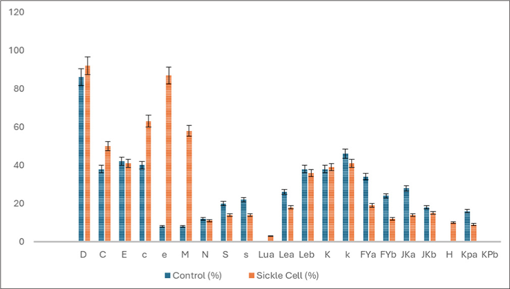
- Frequency, presence and type of minor red cell antigens present in sickle cell disease (SCD) individuals and controls.
| Red cell antigen | Positive (%) | Negative (%) |
| D | 92 (92) | 8 (8) |
| C | 50 (50) | 50 (50) |
| E | 41 (41) | 41 (41) |
| c | 63 (63) | 37 (37) |
| e | 87 (87) | 13 (13) |
| M | 58 (58) | 42 (42) |
| N | 11 (11) | 89 (89) |
| S | 14 (14) | 86 (86) |
| s | 14 (14) | 86 (86) |
| Lua | 3 (3) | 97 (97) |
| Lea | 18 (18) | 82 (82) |
| Leb | 36 (36) | 64 (64) |
| K | 39 (39) | 61 (61) |
| k | 41 (41) | 59 (59) |
| Fya | 19 (19) | 81 (81) |
| Fyb | 12 (12) | 88 (88) |
| JKa | 14 (14) | 86 (86) |
| JKb | 15 (15) | 85 (85) |
| H | 10 (10) | 90 (90) |
| Kpa | 9 (9) | 91 (91) |
| KPb | 0 | 100 (100) |
SCD: Sickle cell disease
DISCUSSION
The age distribution of SCD individuals revealed that individuals in the steady state aged <1 and 10 years had the highest frequency of SCD, although there was no association between age and SCD incidence. SCD across different age groups can be influenced by disease onset and progression, treatment and management considerations, mortality, and survival rates. Usman et al. indicated that most of the participants were aged five;[13] this is consistent with the findings of the present study, possibly because SCD symptoms manifest early in life, typically within the first few months after birth. Infants with SCD may experience complications such as painful vaso-occlusive crises, hand and foot swellings (dactylitis), acute chest disease, and infections, which can lead to hospitalization and medical intervention.[14] 95% of the study population with SCD were homozygous for sickle-hemoglobin (SS), and 1% of the population had CC variants, whereas 4% of the study population were heterozygous for sickle-hemoglobin (SC) variants. Similarly, Nwobuko observed trends in southeastern Nigeria: 98.55% and 1.45% for homozygous sickle-hemoglobin (SS) and heterozygous sickle-hemoglobin C (SC) variants, respectively.[5] There was a noticeable change in sex distribution compared to that of the control and steady-state groups. The dominance of females in the control group and in the steady state may suggest variations in the increase in mortality and morbidity in male SCD individuals, even though SCD is not sex linked.[15] Furthermore, Masese et al. and Fitzmaurice reported that higher female survival may be due to higher fetal hemoglobin levels.[16,17] Understanding the reasons behind the observed shifts could provide insights into how different genders respond during crises. The increase in male representation during the crisis may be indicative of a specific response or vulnerability that was more pronounced in males during crisis scenarios, and this aligns with the work done by Ugwu in Ozalla, the eastern part of Nigeria.[7] Assessing the gender distribution in each group can contribute to a broader understanding of diversity and inclusion dynamics within the community or context under study. The distribution of males and females within each group is likely due to random variation and is not reflective of a systematic association or effect. Further exploration of the factors contributing to these sex distributions could provide a more comprehensive understanding of the observed patterns. In his study in Sokoto, Usman et al. found gender to be equal.[13]
In this study, the prevalence of red cell alloimmunization among multiply transfused SCD individuals in Abuja was 25%, while in Ghana, Boateng et al. reported an alloimmunization prevalence of 16%.[18] Similarly, Natukunda et al. in Uganda documented a lower rate of 6.1%.[19] The prevalence in Cameroon was 55%, while Morocco recorded 90.4% in 2013, and Cote d’ivoire recorded a prevalence of 28.6%, New Delhi (India) recorded a prevalence of 5.2%. By comparison, studies in developed countries such as the United States have reported alloimmunization rates ranging from 18% to 36% in SCD populations, with anti-E, anti-C, and anti-K as the leading specificities.
The results of the alloantibodies observed in this study are suggestive that a subgroup of individuals within the population has developed antibodies against the D antigen, which is a common target for immunization due to its high immunogenicity.[20] The presence of anti-E antibodies in 32% of the population indicates that a larger proportion of individuals have developed antibodies against the E antigen. Anti-E antibodies can pose challenges in transfusion compatibility, as they can lead to adverse reactions if individuals are mismatched with inappropriate blood types. Rh antibodies contributed to approximately 52% of the total antibodies identified, and a total of 24% of the studied population developed anti-Fya antibodies. This antibody development is less common than that of anti-D and anti-E antibodies. Anti-Fya antibodies are specific to the Duffy blood group system and can influence the compatibility of blood transfusions. The development of antibodies against the JKb, Lua, M, Lea, and C antigens in only 4% of the population suggests a decreased prevalence of these specific antibodies. These findings indicate that these antigens may be less immunogenic or less likely to stimulate the production of antibodies in the studied population. The presence of anti-S antibodies was 2% in the population, suggesting a relatively low prevalence. Anti-S antibodies are associated with the S antigen, which is part of the MNSs blood group system. Awareness of the presence of anti-S antibodies is crucial for safe blood transfusion practices. No antibodies were detected in the control group. The most frequent antibodies reported by Adewoyin et al. were anti-E, followed by anti-C before anti-D, which is consistent with the findings of the present study.[21] Anti-D was followed by anti-C, whereas in our study, we observed that anti-Fya was the next most common after anti-D. Wilson et al. reported that the most frequent antibodies were against the E antigen, which is consistent with what we observed from this work.[22] Similarly, Samarah et al. reported the frequency of RBC alloimmunization among SCD individuals to be 7.76%, with anti-K showing the highest frequency.[23] This finding aligns with our findings, but we did not observe anti-K. Coleman et al. reported that 26% of the delay in the hemolytic transfusion reaction was associated with Rh antibodies, while 38% of the non-Rh antibodies were associated with Rh antibodies.[24] In contrast, we identified 52% of the Rh antibodies and 48% of the non-Rh antibodies. Our findings do not agree with those of Coleman et al., who found 26% Rh antibodies and 38% non-Rh antibodies. This could be due to discrepancies in population differences, antigen distribution, reagent sensitivity, or transfusion practices. Kangiwa et al. reported a frequency of alloimmunization among individuals with multiple transfusions of SCD that was consistent with our findings in this study, except for anti-KEL, which was not observed in this study.[25]
These findings in this study align with previous African and Middle Eastern studies, where Rh and Duffy system antibodies are more common due to mismatches in transfused units. However, the absence of anti-K or anti-KEL may be because of the low frequency of the Kell antigen in the donor population, limited antigen exposure in this cohort, or insufficient panel sensitivity for detecting Kell-group antibody antigens.
Ugwu reported that the prevalence of RBC alloantibodies among multiple transfusion individuals with SCD was 9.3%. The antibodies they identified were slightly different from those recorded in the present study, and no antibodies were detected among the control group.[7] Understanding the prevalence of these antibodies within a population of SCD individuals who have been transfused multiple times is vital for proper blood typing and cross-matching to ensure safe and compatible blood transfusions. These findings emphasize the importance of regular antibody screening and careful selection of compatible blood units for transfusion recipients.
The O+ blood group made up a greater percentage of the total number of SCD individuals studied. This finding suggested a greater incidence of SCD among individuals with O+ blood. The A+ and B+ blood groups represented 15% and 19%, respectively. No AB blood group was found in this study. Rh-negative blood groups (O− and B−) accounted for a smaller percentage of the population, with O− (7%) and B− (2%) exhibiting Rh negativity (8%), and they were less common in the SCD population studied. This finding is in line with previous records showing that blood group A had the lowest percentage, group B was a bit higher than group A, group O had the highest prevalence, and the AB blood group was not present.[26] They also recorded a very high Rh positivity and very low percentage of Rh negativity,[27-29] and all the authors also recorded low Rh negativity frequencies in Nigeria. Other researchers have also recorded low frequencies.[30-32] In addition, Ab et al. recorded a high frequency of blood group O in Abuja.[33] The authors equally observed that Rh-positive individuals had the same frequency, and AB- and Rh-negative individuals had the lowest frequency. A high prevalence of blood group O+ was also observed by Olayanju et al.[34] Olayanju et al.[34] and Abbas et al.[35] recorded ABO blood group incidences of O > A > B > AB, while the prevalence of Rh+ was high, which could be influenced by genetic diversity,[36] evolutionary factors, population mixing, and historical dynamics. The specific combination of these factors may have contributed to the high prevalence of both group O and Rh+ blood types within the Nigerian population and Abuja in particular. The frequencies of minor red cell antigen phenotypes in the study population align with the findings of Boateng et al. and Yu et al.[37,38] Lamba et al. found Rhe to be the most common, followed by C, c, and E.[39] This study recorded a frequency of Lea antigen of 18%, which was consistent with the work of Yu et al.[38] For Kpa, 9% was recorded in this study, which was in line with the work of Tariq et al., who reported a low frequency of Lua, which aligns with our work.[27] This could be because certain blood group antigens may exhibit lower frequencies in populations that have been historically isolated. If the Lua antigen is less common in ancestral populations, limited genetic interchange with other populations may have contributed to its low frequency within specific ethnic or regional groups. Within the MNS blood group system, the antigens identified in this study were M (58%), N (11%), S (14%), and S (14%), whereas the occurrence rate of alloimmunization to red cell antigens is a serious complication of transfusion. SCD individuals are prone to developing alloantibodies despite being of the same race as the donor.
Buhari et al. reported a higher frequency among pregnant women in Sokoto, northern Nigeria; however, these antigens were not the most common antigens observed in the population. Within the Duffy blood group system, the antigen frequencies were 19% for Fya and 12% for Fyb, which are higher than those observed by Buhari et al. The Kidd blood group system was found to be moderately low at 14% and 15%, respectively, which is consistent with what Buhari et al. reported and disagrees with what Gwaram and Yusuf reported.[40,41]
These findings underscore several critical considerations for transfusion policy and immunohematologic research in SCD management.
On the Absence of Certain Alloantibodies
Although antibodies such as anti-K, anti-Kpa, and anti-Jkb were not detected in this study population, their absence does not preclude their clinical significance. The lack of these antibodies may reflect the relatively low antigenic exposure within the local donor pool or the sample size limitations of this cohort. Given their known immunogenic potential and association with DHTRs, ongoing immune-hematologic surveillance using larger and more diverse populations is warranted. This would help clarify the prevalence and impact of less commonly detected antibodies in the Nigerian SCD population.
On Blood Donor Registry Development
These findings support the urgent need to enhance Nigeria’s national blood donor registry with extended red cell antigen profiling. Establishing a comprehensive donor database that includes clinically significant antigens—such as Rh subtypes (C, c, E, e), Kell, Duffy, and Kidd—would facilitate better-matched transfusions for patients with SCD. By improving donor-recipient compatibility, such registries can minimize alloimmunization risk, improve transfusion outcomes, and contribute to long-term disease management strategies.
On Extended Matching and Molecular Phenotyping
Standard ABO and RhD matching alone is insufficient for managing transfusion-dependent patients such as those with SCD. The implementation of extended matching policies that incorporate additional antigens such as C, E, K, Fya, and Jkb can significantly reduce alloantibody development. Furthermore, molecular red cell genotyping presents an advanced tool for identifying variant antigens and silent alleles, particularly in multiply transfused patients where serologic typing may be unreliable. Incorporating molecular methods into routine pre-transfusion testing could revolutionize transfusion safety and precision in resource-limited settings.
Summary of findings
The study determined the distribution of minor red cell antigens among multiply transfused SCD individuals and control subjects, and assessed the prevalence and specificity of red cell alloantibodies in the SCD population.
Distribution of minor red cell antigens
The frequency of clinically significant red cell antigens varied among the study population. The Rh (C, c, E, e) and Kell systems were the most represented. For example, RhD positivity was observed in 92%, while K antigen was present in 39% of SCD individuals. These distributions provide important insight into antigen mismatches between donors and recipients, which could predispose to alloimmunization.
Prevalence and specificity of alloantibodies
Alloantibodies were detected in 25% of multiply transfused SCD individuals, confirming the immunogenic risk associated with repeated transfusion. The most common antibodies identified were anti-E, anti-FYa, anti-D, anti-S, anti-C, anti-Lea, anti-M, anti-Lua, and anti-JKb, aligning with known immunogenic targets in transfused SCD populations. No alloantibodies were detected in the control group.
Subgroup trends
Older age groups and female patients showed slightly higher alloantibody rates, although not all differences reached statistical significance. These patterns suggest that both biological and exposure-related factors may influence alloimmunization risk.
Strength of the study
- The study has the potential to aid in the development of more personalized transfusion strategies for SCD individuals, minimizing complications and improving outcomes.
- Conducting this study among a cohort in Abuja allows for a more specific focus on the Nigerian population, which is significant since SCD prevalence is high in Abuja, a cosmopolitan city.
- The research is clinically valuable as it could improve transfusion compatibility and reduce the risk of adverse reactions.
Limitations of the Study
- A longitudinal (cohort study) approach would provide a better understanding of how these parameters change over time and with repeated transfusions, offering more robust insights into the long-term risks and immune responses of SCD individuals, this may be considered as further work.
- Variability in laboratory techniques or limited access to advanced testing facilities could affect the accuracy and reproducibility of the findings.
Cautious Interpretation of Results
The findings highlight a moderate prevalence of clinically significant alloantibodies, with anti-E, anti-FYa, anti-D, anti-S, anti-C, anti-Lea, anti-M, anti-Lua, and anti-JKb emerging as the most commonly encountered specificities. These results align with prior studies in similar settings, reinforcing the need for more antigen-matched transfusion practices for individuals with SCD. The data also show that alloimmunization risk increases with the number of transfusions, supporting a dose-dependent relationship between antigenic exposure and immune sensitization. While trends suggesting higher alloimmunization rates among older and female patients were noted, these did not consistently reach statistical significance and should be interpreted with caution. These subgroup findings, though suggestive, would benefit from further exploration in larger cohorts. Importantly, the interpretation of our results must take into account several limitations. First, the use of purposive sampling and the relatively small sample size may limit the generalizability of the findings to the broader SCD population in Nigeria. Second, some transfusion histories were based on patient recall or incomplete records, introducing the potential for recall bias or misclassification. Third, the cross-sectional design precludes any inference of causality between transfusion burden and alloantibody formation. The multiplicity of analyses, including subgroup and sensitivity analyses, raises the possibility of type I error, especially in the absence of adjustment for multiple comparisons. Nonetheless, these analyses were exploratory in nature and aimed to guide future hypothesis generation. When compared with evidence from other low-resource settings, our findings are consistent with reported alloantibody prevalence rates and specificities. However, variations in donor pool composition, antigenic exposure, and laboratory methods across studies underscore the importance of local data to inform transfusion policy.
Generalizability (External Validity) of the Study Results
The generalizability of our findings is subject to certain constraints due to the study design and setting. The use of purposive sampling and the selection of tertiary healthcare institutions within Abuja means that the sample may not fully represent all individuals living with SCD across Nigeria, particularly those in rural or underserved regions. Differences in transfusion practices, access to healthcare, and the composition of the donor pool in other parts of the country may affect the applicability of our results elsewhere.
Moreover, since the study population consisted of patients receiving care at well-resourced tertiary centers, the observed prevalence of alloantibodies and red cell antigen distribution might differ from those in primary or secondary healthcare settings where transfusion monitoring may be less robust. Therefore, extrapolation of the findings to the national level or to other low-resource settings should be done cautiously.
Nonetheless, the consistency of our results with those from similar studies conducted in other sub-Saharan African countries supports a degree of external validity. The patterns of alloantibody specificity, particularly the predominance of anti-E, anti-FYa, anti-D, anti-S, anti-C, anti-Lea, anti-M, anti-Lua, and anti-JKb, align with existing literature and suggest that the immunologic risks associated with transfusion in SCD patients are broadly similar across comparable settings.
Implications for national blood policies
Adopt SCD-specific matching standards
- Move from ABO/RhD-only to prophylactic extended matching at least for Rh (C/c, E/e) and Kell (K) for all SCD patients; extend to Duffy/Kidd/MNS for patients with prior alloimmunization.
- Prioritize pre-storage leukoreduction for all SCD units to reduce febrile reactions and CMV transmission.
Scale red-cell phenotyping/genotyping
- Baseline extended phenotype (or genotype) for every SCD patient before the first transfusion; repeat after any transfusion reaction.
- Build regional reference labs (e.g., Abuja as a hub) with molecular genotyping panels optimized for African RHD/ RHCE variants; link results to the electronic blood bank.
Targeted donor recruitment to match patient antigen profiles
- Recruit and retain voluntary, repeat donors from the same ethnolinguistic backgrounds as SCD patients to improve availability of Rh/K-matched units.
- Create a “phenotyped donor registry” (flagging rare phenotypes and K-negative/Rh-variant donors) with rapid recall capability.
National hemovigilance and data systems
- Mandate reporting of alloantibody formation, DHTRs, and hyperhemolysis using a standardized national form.
- Use data to forecast demand for antigen-negative units and to monitor the impact of matching policies.
Standardized transfusion indications and pathways
- National guideline for simple versus exchange transfusion (acute chest syndrome, stroke, pre-operative, severe anemia), with restrictive thresholds where safe to limit exposure.
- Transfusion passports for patients (history of antibodies, special requirements) and a 24/7 consult line at reference centers.
Supply chain and financing
- Ring-fence funding (NHIA/Basic Health Care Provision Fund) for reagents, genotyping kits, leukoreduction filters, and staff training.
- Contracting and pooled procurement through the National Blood Service Commission to lower costs and standardize quality.
Training and accreditation
- Mandatory competency training for blood bank and clinical staff on SCD transfusion risks, antibody workups, and DHTR management.
- Accredit blood banks based on their ability to provide extended-matched units to SCD patients.
CONCLUSION
The findings of this work showed that 25% of SCD individuals developed alloantibodies, and anti-E was the most common, followed by anti-Fya and anti-D, while all the controls were non-reactive. The distribution of minor RBC antigens in sickle cell individuals showed that the D antigen was more frequent than antigen e and C in SCD individuals attending tertiary health care centers in Abuja. Identifying alloantibodies in SCD individuals is crucial, as it can have implications for future transfusions and impact the overall management of the disease.
Consent to Participate
Both written and verbal informed consent and assent were obtained from individuals and parents of minors who volunteered for this study.
Data Availability Statement
The datasets used and analyzed during the current study are available from the corresponding author on reasonable request.
Key Messages
(1) A measurable prevalence of red cell alloantibodies was identified among multiply transfused SCD patients in selected tertiary institutions in Abuja, confirming alloimmunization as a relevant transfusion-related complication in this population. (2) Alloantibodies detected were predominantly directed against clinically significant blood group systems, particularly Rh, consistent with the observed distribution of red cell antigens among the study participants. (3)Increasing transfusion exposure was associated with a higher likelihood of alloantibody formation, supporting the need for routine antibody screening and extended red cell antigen matching in the transfusion management of SCD patients.
Acknowledgments:
The authors wish to acknowledge the efforts and assistance given by the members of the staff of the Sickle cell clinics at all the sites used for this study and the contribution of Prof. Olayanju AO Kingston University, UK.
COMPLIANCE WITH ETHICAL STANDARDS
Conflicts of Interest: Not applicable. Financial Disclosure: Not applicable. Funding/Support: Not applicable. Ethics Approval: This study was conducted in accordance with the 1964 Declaration of Helsinki. Ethical approvals were obtained from the Health Research Ethics Committee of the College of Medicine and Health Sciences, Afe Babalola University, Ado Ekiti, Ekiti State, Nigeria (ABUADHREC/12/06/2021/056); National Hospital, Abuja, Nigeria (NHA/EC/083/2022); Federal Medical Center Jabi, Abuja, Nigeria (FMCABJREC/ HREC/2022/076); and University of Abuja Teaching Hospital, Gwagwalada, Abuja, Nigeria (FCT/UATH/HREC/1085). Declaration of Patient Consent: Consent obtained from all participants and parents/guardians of minors. Use of Artificial Intelligence (AI)-Assisted Technology for Manuscript Preparation: The authors confirm that they have used artificial intelligence (AI)-assisted technology solely for language refinement and to improve the clarity of writing. No AI assistance was employed in the generation of scientific content, data analysis or interpretation. Disclaimer: None.
REFERENCES
- Defining global strategies to improve outcomes in sickle cell disease: A Lancet Haematology Commission. Lancet Hematol.2023 Aug 01;10(8):e633-86.
- [CrossRef] [PubMed] [Google Scholar]
- Paediatric acute respiratory distress syndrome incidence and epidemiology (PARDIE): An international, observational study.Lancet Respir Med. 2019
Feb ;7(2):115-28.- [Google Scholar]
- Genotypic and phenotypic composition of sickle cell disease in the Arab population - a systematic review.Pharmgenomics Pers Med. 2023
Feb 21 ;16:133-44.- [CrossRef] [PubMed] [Google Scholar]
- Gender-related differences in sickle cell disease in a pediatric cohort: A single-center retrospective study.Front Mol Biosci.
2019 Dec 05 ;6:140.- [CrossRef] [PubMed] [Google Scholar]
- Sex-based differences in the manifestations and complications of sickle cell disease: Report from the sickle cell disease implementation Consortium.PLoS One. 2021
Oct 29 ;16(10):e0258638.- [CrossRef] [PubMed] [Google Scholar]
- One third of alloantibodies in patients with sickle cell disease transfused with African blood are missed by the standard red blood cell test panel. Haematologica.2021 Aug 01;106(8):2274-6.
- [CrossRef] [PubMed] [Google Scholar]
- Red blood cell alloimmunization and minor red blood cell antigen phenotypes in transfused Ghanaian patients with sickle cell disease.Transfusion. 2019 Jun;59(6):2016-22.
- [CrossRef] [PubMed] [Google Scholar]
- Frequencies of red blood cell major blood group antigens and phenotypes in the Chinese Han population from Mainland China.Int J Immunogenet. 2016 Aug;43(4):226-35.
- [CrossRef] [PubMed] [Google Scholar]